
Stratasys and Siemens Healthineers have developed 3D printed, patient-specific anatomical models that replicate human tissue with incredible accuracy, transforming medical imaging, surgical planning, and education.
Traditionally, surgeons have relied on generic models or cadavers for practice, which often don’t capture the unique complexity of an individual patient’s anatomy. This can lead to unforeseen challenges during actual procedures since real patients often have anatomical variations that the standard training methods don’t incorporate. Studies have shown that misinterpretation of anatomical structures is a key risk factor for surgical complications, such as bile duct injuries in gallbladder surgeries.
Additionally, cadavers lack living tissue properties and rarely represent the exact pathology of a given patient, making it difficult for surgeons to anticipate case-specific challenges. Even advanced imaging, like CT or MRI scans, doesn’t always provide a full 3D understanding of complex structures, leading to unforeseen difficulties once surgery begins. Polish researchers published an article in 2022 that indicates that many surgeons recognize the limitations of traditional training, with 76% highlighting the importance of understanding anatomical variations to avoid complications in surgery.
To tackle these issues, surgeons turned to 3D printed anatomical models and virtual reality (VR) simulations, allowing them to train using patient-specific data, improving preparation and outcomes. In fact, using 3D printing to create exact replicas of a patient’s anatomy has been linked to reduced surgical time by an average of 62 minutes per case and fewer intraoperative errors, according to researchers at Washington University School of Medicine. These training tools help surgeons anticipate and navigate complex anatomical structures before entering the operating room.
Recognizing the need for even more precise anatomical models, Stratasys partnered with Siemens’ subsidiary Siemens Healthineers to pioneer a solution for pre-surgical planning and medical education.
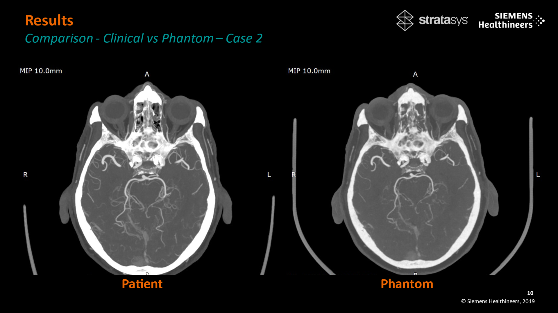
Patient-specific 3D-printed Phantom. Image courtesy of Stratasys.
By harnessing Stratasys’ proprietary RadioMatrix materials and Digital Anatomy technology, the partnership has made it possible to create 3D printed models that mirror the exact anatomical structures of patients. RadioMatrix materials allow these models to appear realistically in CT scans, while Digital Anatomy technology recreates the texture and density of real human tissue. Additionally, Siemens Healthineers’ advanced imaging algorithms help refine these models, ensuring they closely match real patient anatomy in structure and radiographic appearance. Overall, these detailed, patient-specific “phantoms” improve imaging accuracy and provide a lifelike feel for surgeons to test them before surgery.
This advance improves medical imaging in several ways. It allows professionals to fine-tune CT scanners with models closely mimicking human anatomy, leading to sharper images and more precise diagnostics. Researchers can also use these accurate replicas to train AI imaging algorithms more effectively, opening the door to new clinical applications.
Moreover, these 3D printed phantoms provide a cheaper alternative to cadavers, making medical training more accessible, sustainable, and ethical. A single cadaver can cost around $2,000, not including expenses for storage and maintenance, while operating an anatomy lab can add up to $17,500 annually. On the other hand, 3D printed models can be made on-demand, cutting sourcing and maintenance costs while offering a reusable, highly detailed alternative for medical education.
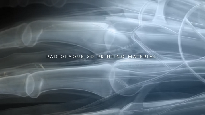
Stratasys radiopaque RadioMatrix material. Image courtesy of Stratasys.
The importance of this collaboration was pointed out at the Radiological Society of North America (RSNA) annual meeting, where the duo showcased their research findings. Research shows that these 3D printed models are incredibly detailed, with small differences from real human tissue that are almost undetectable on CT scans. The accuracy is measured in Hounsfield units (HU), the scale used to determine how different tissues appear in medical imaging. The variations in critical areas like grey matter and veins are as low as a single HU, making these models highly reliable for detailed medical imaging and surgical planning.
Jesús Fernández Léon, Head of Computed Tomography Product & Clinical Marketing at Siemens Healthineers, said “The integration of 3D-printing solutions to create patient-realistic CT phantoms, combined with the Digital Anatomy technology from Stratasys, represents a significant innovation in the field of computed tomography. This cooperation not only enhances our ability to assess and verify the performance of modern CT systems but also ensures that our algorithms can rely on a highly realistic depiction of human anatomy. By working together, we are setting new standards in medical imaging.”
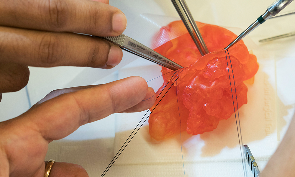
Stratasys J750 Digital Anatomy can help standardize surgical skills and delivery of care by practicing on the most accurate representation of the targeted pathology. Image courtesy of Stratasys
Beyond improving imaging and surgical planning, this technology is set to transform multiple areas of healthcare. For example, radiologists can interpret scans more confidently, leading to more precise diagnoses. The realistic nature of these 3D printed phantoms also supports the development and testing of AI-based diagnostic tools, which could help detect diseases earlier. Additionally, they offer a valuable training resource for medical students and professionals, allowing them to practice with lifelike models that copy human tissues and prepare better for real surgeries.
According to Stratasys Healthcare VP Erez Ben Zvi, this collaboration will pave the way for innovations that enhance imaging precision, improve training efficiency, and reduce reliance on cadavers.
While cadaver-based teaching has long been the main method in medical education, its drawbacks are becoming clearer. A 2018 study by the Institute of Anatomy at Medical School Brandenburg analyzing 71 countries found that cadaver-based teaching remains dominant in medical schools worldwide, with 68 countries incorporating it into their curricula—32% relying solely on body donations, 31% using unclaimed bodies, and the rest using a mix of both.
However, the use of 3D printed anatomical models is gaining traction, particularly in the United States and Australia, as highlighted in a Newcastle University 2023 study, which notes the increasing adoption of 3D printing to enhance or, in some cases, replace traditional cadaveric dissection in medical education.
“This is a game-changer for the medical community. We believe this work can speed up the advancement of medicine and improve patient outcomes,” said Ben Zvi.
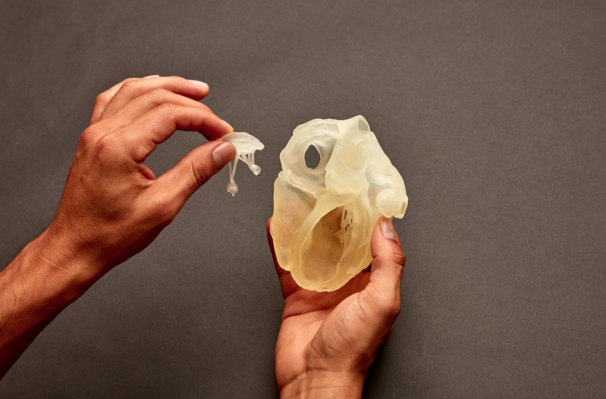
Medical 3D printing with Stratasys’ J750 Digital Anatomy printer. Image courtesy of Stratasys.
Stratasys states that integrating 3D printing with advanced imaging marks a major shift in medical practice. As hospitals and imaging centers adopt patient-specific phantoms, surgical planning becomes more precise, reducing risks and improving recovery times. These models also speed up medical research, making testing and approving new devices and procedures easier. Also, they hope to provide medical institutions worldwide with accessible, high-quality training tools without the ethical and logistical challenges of retrieving cadavers.
Subscribe to Our Email Newsletter
Stay up-to-date on all the latest news from the 3D printing industry and receive information and offers from third party vendors.

