
Researchers at the University of California, Irvine (UC Irvine), and the New York Institute of Technology have 3D printed one of the most realistic models of the human colon ever made. The advance could change how we study colon cancer and test new drugs.
The team used bioprinting techniques to create a miniature, life-like colon from human cells. Unlike flat petri-dish cultures or animal tests, this 3D printed “human colon-on-a-lab” mimics the real shape, texture, and behavior of the organ, including its natural folds and tissue layers, explained the researchers in their paper “Development of a 3D Human Colon Model Along with Bioelectronics for the Study of Colorectal Cancer and Drug Response,” published in the journal Advanced Science.
They call their creation the 3D-IVM-HC model, short for 3D In Vitro Model of the Human Colon, a bioprinted system that combines living human cells with built-in bioelectronic sensors to monitor tissue health in real time.
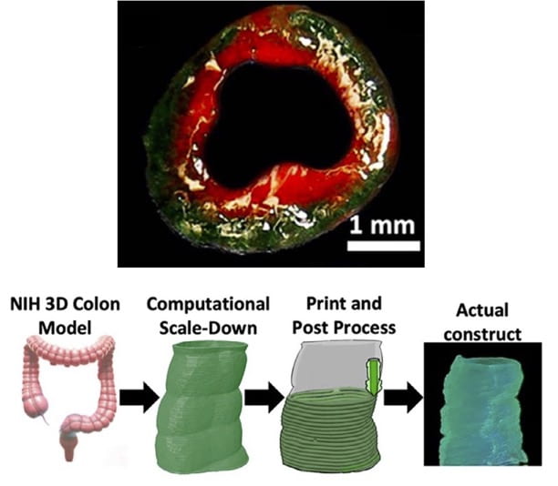
Design and biofabrication of the 3D-IVM-HC model. Image courtesy of UC Irvine.
The model was printed using a technique called FRESH (Freeform Reversible Embedding of Suspended Hydrogels), originally developed by Adam Feinberg’s lab at Carnegie Mellon University in 2015. FRESH is a cornerstone method in modern bioprinting because it solves one of the field’s biggest challenges: printing soft, delicate biological materials that normally collapses under their own weight. It works by extruding these materials into a supportive gel bath that temporarily holds the structure in place as it solidifies, allowing scientists to 3D print complex, freeform shapes like tissues and organs with fine internal details.
The foundational IP for FRESH is exclusively licensed to Feinberg’s spinout FluidForm Bio, and the method has been widely adopted in academic bioprinting. With more than 2,000 citations, the original Science Advances paper ranks among the most influential publications in bioprinting, a rare achievement in a field where most studies receive fewer than 50 citations.
In this particular study, the researchers used FRESH to 3D print a miniature colon about 5 by 10 millimeters in size. They combined gelatin methacrylate (GelMA) and alginate to create a flexible matrix that mimics the feel and function of living tissue, which is strong enough to maintain form, but soft enough for human cells to thrive inside.
After printing, they seeded the model with two types of cells: fibroblasts (which make up the colon’s connective tissue) and Caco-2 epithelial cells (which line the inside of the colon). Over time, these cells organized themselves into layers and formed “crypt-like structures,” which are the tiny folds that increase surface area inside the real human colon, explain the researchers. According to the study, the printed tissue showed over 95% cell survival and realistic tissue stiffness, making it far more accurate than traditional lab cultures.
To test how realistic the model was, the researchers inserted colon cancer cells (HCT116) and treated them with 5-fluorouracil (5-FU), a common chemotherapy drug. In standard 2D lab tests, cancer cells responded strongly to the drug, but in the 3D printed colon, they showed much higher resistance, just like in real patients. This means the 3D model may predict clinical outcomes more accurately than flat cell cultures or animal tests.
According to senior author Rahim Esfandyar-pour, UC Irvine assistant professor of electrical engineering and computer science, the model’s precise 3D structure is central to recreating the mechanical and chemical environment that governs real cellular behavior, something that flat cultures or even some animal models can’t replicate.
The team also integrated bioelectronic sensors into the system to monitor electrical resistance (TEER), a measure of how well the tissue forms a barrier, something critical for testing how drugs and diseases affect the colon’s lining. The printed tissue achieved TEER values close to those of real human samples, confirming that it behaves like living tissue.
The team noted that roughly half of the toxicology results from rodent experiments fail to translate effectively to humans, and animal studies can take years and cost millions. By contrast, the 3D-IVM-HC model can be developed in about two weeks and used for drug testing within days.
According to lead authors Jorge Alfonso Tavares-Negrete and Rahim Esfandyarpour, the 3D printed colon offers a faster, cheaper, and more ethical way to study human disease, cutting animal testing costs by up to 80% and providing a realistic, customizable platform for testing cancer treatments.
“By utilizing our human-cell-based, animal-free approach, the 3D-IVM-HC model substantially reduces experimental costs (by roughly 70–80%, required for animal procurement, housing, and regulatory compliance) and streamlines timelines, thus facilitating rapid, cost-effective, more ethical, and scalable translational studies. Importantly, eliminating interspecies variability in our 3D-IVM-HC model is expected to enhance clinical translatability, providing an accelerated and ethically responsible pathway for preclinical research,” states the study.
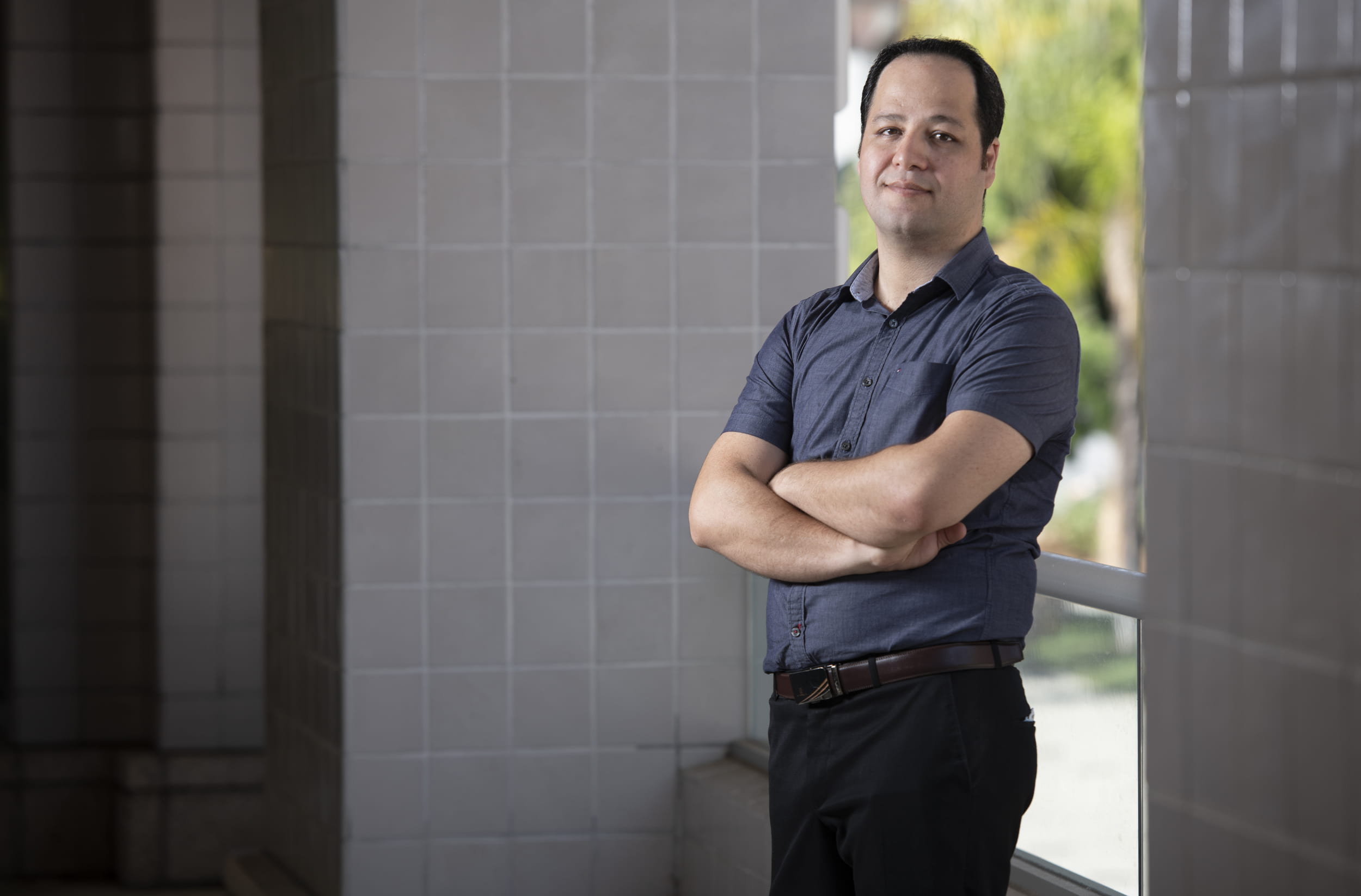
UC Irvine Professor Rahim Esfandyar-pour. Image courtesy of Steve Zylius / UC Irvine.
The researchers believe this model could become a powerful new tool for colorectal cancer research, drug screening, and personalized medicine. It may also pave the way for 3D printed models of other organs, bringing us closer to lab-grown systems that truly reflect the human body.
“Hospitals and laboratories could ultimately use such models to run preclinical tests on new therapies in an ethical, timely manner, possibly transforming the drug development pipeline. This research may represent a significant step toward global efforts to develop more reliable, humane and cost-effective alternatives to animal testing, potentially advancing precision medicine and improving outcomes for patients with colorectal cancer worldwide,” stated Esfandyar-pour.
Looking ahead, the team envisions using patient-derived cells to create personalized mini-colons for testing how an individual’s tumor responds to specific drugs, a step toward true precision medicine.
Subscribe to Our Email Newsletter
Stay up-to-date on all the latest news from the 3D printing industry and receive information and offers from third party vendors.
Upload your 3D Models and get them printed quickly and efficiently.


